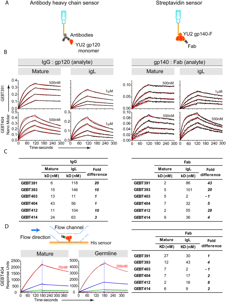
Figure 3. igL and mature antibody binding kinetics to gp120 monomer and gp140-F immunogen.
(A) Schematic of the BLI assay formats. IgGs were captured on anti-human IgG Fc BLI biosensors and monomeric gp120, the analyte in solution and gp140-F was captured on streptavidin biosensors with the analyte in solution being Fabs. (B) Binding curves of selected igL IgG and their corresponding mature mAbs to SEC-purified monomeric YU2 gp120 (left) and to YU2 gp140-F (right). Data points are shown in black and the corresponding fits are shown in red. The Env concentrations used ranged from 500 nM to 1μM initial concentration as indicated above the highest concentration curve. 1:2 serial dilutions were used subsequently. Mature antibodies in general have higher affinity to gp120, displaying faster on-rates and slower off-rates. (C) Binding kinetics of the mature and igL IgG and Fabs to YU2 gp120 and YU2 gp140-F trimers respectively. (D) SPR binding kinetics were determined and used to derive affinities of both the mature and igL Fabs, with fold differences indicated (right), and the schematic of the method and binding curves are shown (left).

