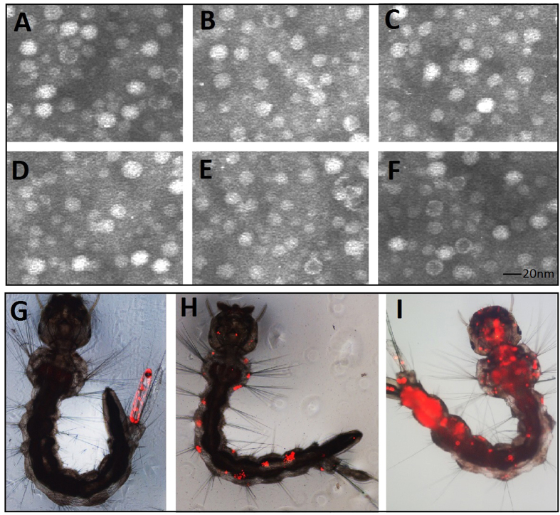
Figure 6. Electron microscopy of the recombinant virus particles from media of infected cultures.
(A–F) Electron micrographs of the virus particles stained with 1% uranyl acetate. All of the recombinant viruses and wild-type viral particles displayed a homogeneous size (approximately 20 nm in diameter). (A) VrepUCA-7, (B) VrepUCA-210, (C) VrepUCA-7s, (D) VrepUCA-210s, (E) VrepUCA-antiV, and (F) AaeDV (recombinant Aedes aegypti densovirus). Primary infection site in Aedes albopictus larvae transduced with red fluorescent protein (DsRed) marker; (G) anal papillae; (H) bristle cell; (I) the whole body distribution of AaeDV from the primary infection site.

