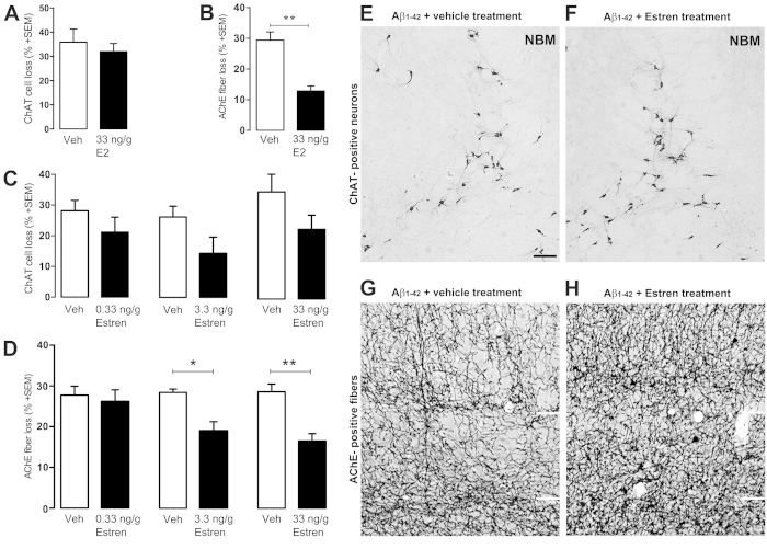
Figure 2. Estren attenuates Aβ1–42–induced lesions of cortical projections.
Aβ1–42 -induced ChAT cell and fiber loss in estradiol (A,B), estren (C–F) treated mice compared to the vehicle treated group. Photomicrographs demonstrate ChAT positive cell bodies in the NBM and AChE-stained fibers in layer IV and V of the somatosensory cortex at the contralateral nonlesioned (E,G) brain side and ipsilateral lesioned (F,H) side after 12 d of Aβ1–42 injection and estren administration. Scale bar, 50 μm. Histograms show mean ± SEM (n = 4–9). *P < 0.05; **P < 0.01; ***P < 0.001 (t-test (A,B) or ANOVA with post hoc Tukey test).

