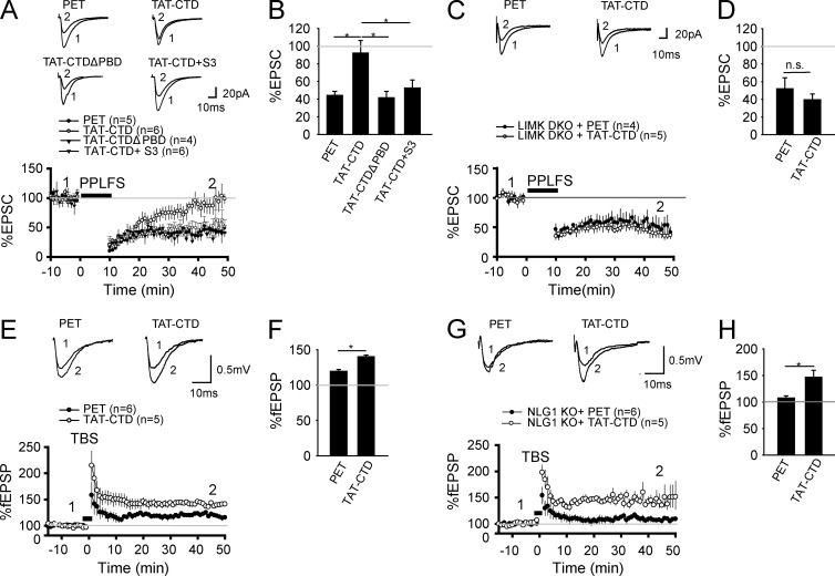
Figure 7.
NLG1 CTD inhibits LTD and facilitates LTP. (A) Whole-cell recordings of CA1 neurons of WT hippocampal slices pretreated with the recombinant proteins showing that TAT-CTD, but not PET or TAT-CTDΔPBD, blocked PP-LFS–induced LTD. TAT-CTD failed to block LTD when the S3 peptide was included in the recording electrode (TAT-CTD + S3). (B) Summary graph of A showing significant differences in PP-LFS–induced LTD between TAT-CTD and PET, TAT-CTDΔPBD, or TAT-CTD + S3–treated slices. (C) Whole-cell recordings of CA1 neurons of LIMK1/2 double-KO hippocampal slices pretreated with TAT-CTD or PET showing that TAT-CTD failed to block PP-LFS–induced LTD in these mice. (D) Summary graph of C showing similar LTD in TAT-CTD– and PET-treated slices of LIMK1/2 double-KO mice. n.s., not significant. (E) Field recordings in the CA1 region of WT hippocampal slices pretreated with TAT-CTD or PET showing that TAT-CTD enhanced TBS-induced LTP compared with PET. (F) Summary graph of E showing significantly higher LTP in TAT-CTD compared with PET-treated slices. (G) Field recordings of NLG1 KO hippocampal slices pretreated with TAT-CTD or PET showing that TAT-CTD enhanced TBS-induced LTP compared with PET. (H) Summary graph of G showing significantly higher LTP in TAT-CTD compared with PET-treated slices. *, P < 0.05.

