Fig. 3.
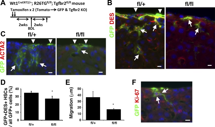
Conditional deletion of Tgfbr2 gene in MCs in biliary fibrosis induced by BDL. A: the tamoxifen-induced CreERT2 in MCs activates expression of GFP, while deleting Tgfbr2 gene in Wt1CreERT2/+;R26TGfl/fl;Tgfbr2fl/fl mice. After deletion of Tgfbr2 gene, biliary fibrosis was induced by bile duct ligation (BDL). B and C: immunofluorescence staining of the liver from the fl/+ and fl/fl mice 2 wk after BDL. The liver tissues were immunostained with antibodies against GFP (green) and DES or ACTA2 (red). Arrowheads and arrows indicate GFP+ MCs and HSCs, respectively. Nuclei were counterstained with DAPI. Bars: 10 μm. D: quantification of GFP+ HSCs in all GFP+ cells in the fl/+ and fl/fl mouse livers after BDL. The Tgfbr2 knockout decreases the percentage of GFP+ myofibroblasts. *P < 0.05 (n = 3 for each group). E: migration of GFP+ DES+ HSCs in all GFP+ cells in the fl/+ and fl/fl mouse livers. F: immunofluorescence staining of fl/+ mouse livers after BDL using antibodies against GFP (green) and Ki-67 (red). Arrows indicate Ki-67+ GFP+ MC-derived HSCs.

