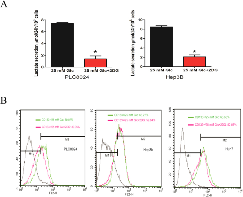
Figure 4.
(A) Lactate was measured fluorometrically. Cells were seeded and serum starved 16 h followed by replaced with fresh medium in presence of 20 mM 2-DG or not. Lactate measured in cell culture supernatants showing quantity (μmol) per 106 of tested cells. Samples were diluted 2–5 fold. (B) HCC cell lines were cultured under high glucose medium in presence of 20 mM 2-DG or vehicle control (DMEM only) for 16 h, and CD133-positive phenotype were detected by flow-cytometry. Each experiment was repeated at least three times. Representative data are shown.

