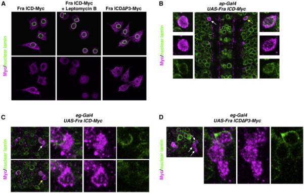
Figure 4. The Fra ICD shuttles between the cytoplasm and the nucleus.

A) S2R+ cells were transfected with the indicated Myc-tagged constructs and treated with Leptomycin B or vehicle, as indicated. Cells were immunostained with antibodies against Myc (magenta) and nuclear lamin (green). For each condition, a single optical plane is shown.
B) Stage 16 embryo in which ap-Gal4 is driving expression of UAS-Fra ICD-Myc. Anterior is up. A single optical plane is shown. The embryo is stained with antibodies against Myc (magenta) and nuclear lamin (green). White arrow indicates a cell in which the Fra ICD is enriched in the nucleus (enlarged in the panels on the left). Yellow arrow indicates a cell in which the Fra ICD is largely excluded from the nucleus (enlarged in the panels on the right).
C) One segment of a stage 14 embryo in which eg-Gal4 is driving expression of UASFra ICD-Myc. Anterior is up. Two different single optical planes are shown. The embryo is stained with antibodies against Myc (magenta) and nuclear lamin (green). In the top row, the white arrow indicates a cell in which the Fra ICD is enriched in the nucleus (enlarged in the panels on the right). In the bottom row, the yellow arrow indicates a cell in which the Fra ICD is largely excluded from the nucleus (enlarged in the panels on the right).
D) One segment of a stage 14 embryo in which eg-Gal4 is driving expression of UASFra ICDΔP3-Myc. Anterior is up. A single optical plane is shown. The embryo is stained with antibodies against Myc (magenta) and nuclear lamin (green). White arrows indicate three cells in which the Fra ICD is enriched in the nucleus (enlarged in the panels on the right).
See also Figure S1.
