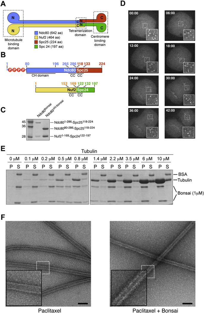
Figure 1. Properties of Ndc80bonsai.
(A) Organization of Ndc80 subunits.
(B) Scheme of Ndc80-Spc25 and Nuf2-Spc24 fusion proteins. Residues 1–286 of Ndc80 (Ndc801–286) were fused to residues 118–224 of Spc25 (Spc25118–224). Residues 1–169 of Nuf2 (Nuf21–169) were fused to residues 122–197 of Spc24 (Spc24122–197). Red circles with “P” mark phosphorylation sites in the Ndc80 N-terminal tail (Cheeseman et al., 2006; DeLuca et al., 2006; Wei et al., 2007).
(C) Ndc80-Spc25 and Nuf2-Spc24 fusions were coexpressed in E. coli and purified to homogeneity.
(D) When injected in HeLa cells, Alexa Fluor 488-labeled Ndc80bonsai stained kinetochores throughout mitosis.
(E) Partition of the Ndc80bonsai complex in pellet (P) and supernatant (S) fractions in cosedimentation assay with increasing concentrations of polymeric tubulin.
(F) Negative stain EM images of Paclitaxel-stabilized microtubules in the absence (left) and presence (right) of bound Ndc80bonsai. A thick halo of protein surrounds the microtubules bound by Ndc80bonsai, giving them a hairy appearance. Insets are 2.5×. Bar = 100 nm.

