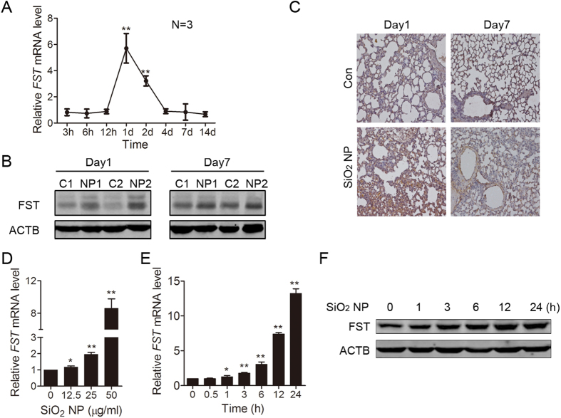
Figure 2. SiO2 NP enhances FST expression in mouse lung tissue and in A549 cells.
(A–C) Mouse lung was instilled with PBS or 100 μg of SiO2 NPs and harvested at the indicated time. (A) The FST mRNA level in mouse lung tissue was measured by real time qPCR and normalized to ACTB mRNA level. (B) The lung tissue protein lysates from paired mice were subjected to immunoblotting for FST detection. Two mice were used for each group (C: PBS; NP: SiO2 NP). (C) Fixed lung tissues from paired mice were subjected to immunohistochemistry with FST antibody. (D) A549 cells were incubated with SiO2 NPs at different concentrations for 12 h. The FST mRNA level was measured by real time qPCR. Data are presented as the mean ± SD of three independent experiments. *P < 0.05, **P < 0.01. (E,F) A549 cells were incubated with or without 50 μg/ml of SiO2 NPs and harvested at the indicated time. (E) The FST mRNA level was measured by real time qPCR. Data are presented as the mean ± SD of three independent experiments. *P < 0.05, **P < 0.01. (F) The FST protein level was detected by immunoblotting.

