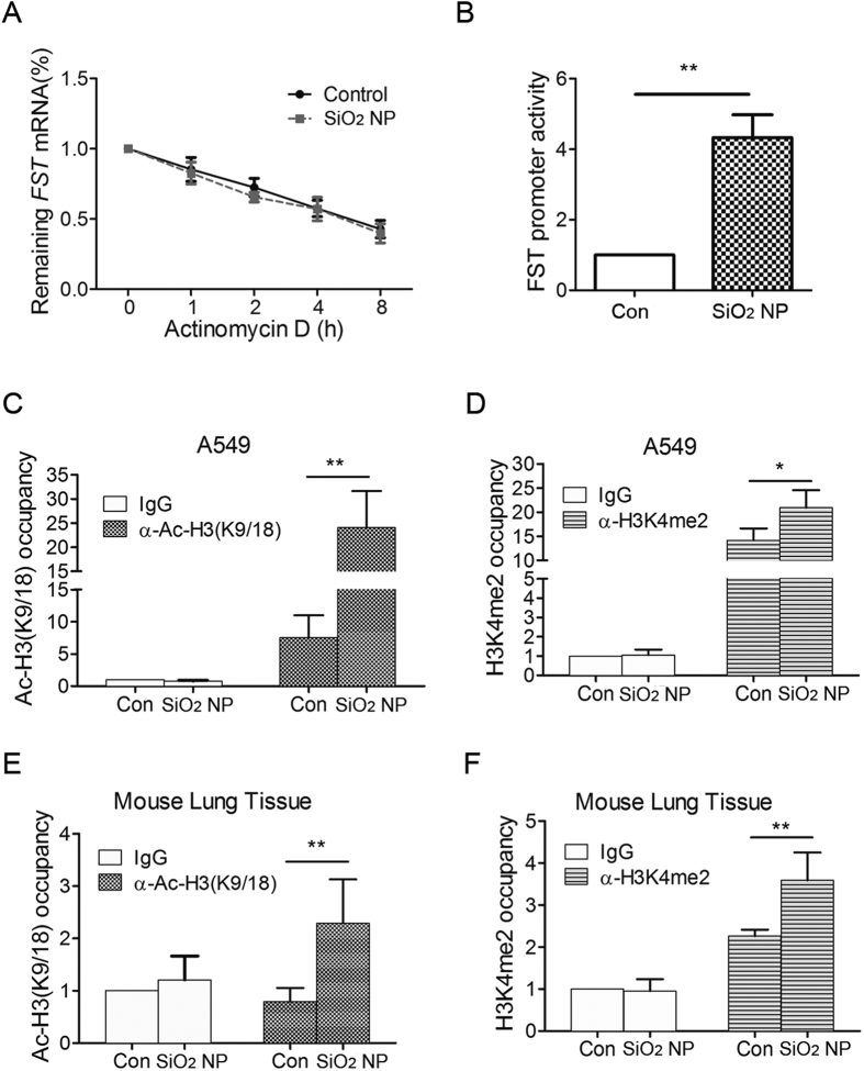
Figure 3. The transcription of FST gene is activated during SiO2 NP treatment.
(A) A549 cells treated with or without SiO2 NPs were incubated with 5 μg/ml of actinomycin D for 1, 2, 4 and 8 h. FST mRNA level was measured and normalized to ACTB mRNA level. (B) A549 cells transfected with FST promoter luciferase reporter and renilla luciferase internal control were treated with or without 50 μg/ml of SiO2 NPs for 12 h. Luciferase activity was then detected and normalized to renilla activity. (C,D) A549 cells were treated with or without 50 μg/ml of SiO2 NPs for 12 h. ChIP analysis was performed with antibodies against Ac-H3(9/18) (C) or H3K4me2 (D) and analyzed by qPCR. (E,F) Mouse lung was instilled with PBS or 100 μg of SiO2 NPs for 1 day. ChIP analysis was performed with antibodies against Ac-H3(9/18) (E) or H3K4me2 (F) and analyzed by qPCR. The occupancies of Ac-H3(9/18) or H3K4me2 at FST promoter region were normalized to ACTB. The values are the mean ± SD of three independent experiments. *P < 0.05, **P < 0.01.

