
Fig. 2.
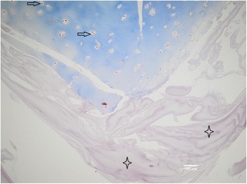
Particulated cartilage (in blue) embedded in fibrin matrix (in violet), ×10 magnification. Trichrome staining is not showing any presence of connective tissue in fibrin matrix (marked with four-point stars). Chondrocytes (marked with arrow) are located in the lacunas; there is no sign of cell migration into the matrix after 5 weeks of cultivation