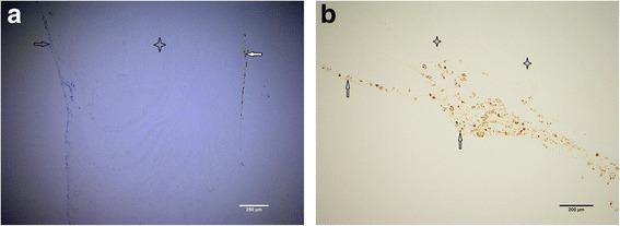
Fig. 4.

a Particulated cartilage fragments (stained in brown to the right with S-100) digested overnight with collagenase solution and thereafter embedded in fibrin matrix (transparent in the middle), ×10 magnification. Bone marrow-originated cells could be visible to the left stained in blue. These were lying at the upper phase of the perforated nylon membrane, which has been removed during the sample preparation. Both chondrocytes (marked with white arrow) and bone marrow-originated cells (marked with black arrow) have attached to the fibrin matrix (marked with four-point star) but have not penetrated it after 5 weeks of cultivation. b Particulated cartilage fragments digested overnight with collagenase solution and thereafter embedded in fibrin, ×10 magnification. Chondrocytes are stained (marked with arrow) with S-100 and have attached to the fibrin matrix (marked with four-point stars) but have not penetrated it after 5 weeks of cultivation
