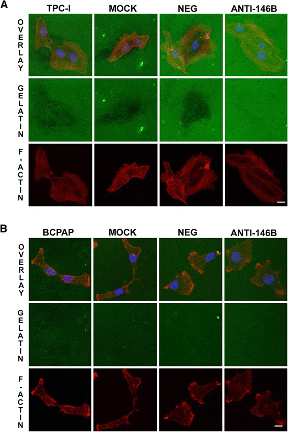
Fig. 3.

Inhibition of miR-146b-5p decreased gelatin degradation by TPC-1 cells. Forty hours after transfection, cells were seeded upon glass coverslips (18 mm) coated with fluorescent gelatin (green) and cultured for 8 h. After this period, cells were fixed, stained for F-actin (red) and nucleus (blue) and analyzed by confocal microscopy. Representative images are shown for TPC- 1 (a) and BCPAP (b) cells. The degradation activity of control and treated groups (miR-146b-5p inhibited) are identified as dark areas on gelatin-FITC background. TPC-1 / BCPAP: cell, Mock: cell + transfection agent, Neg: cell + anti-miR negative control, Anti-146b: cell + anti-miR-146b
