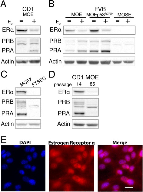
Fig. 1.

Receptor status and estrogen responsiveness monitored by Western blot analysis. a Analysis of ERα and PR expression in response to 24 h 17β-estradiol (1nM, E2) treatment in CD1 MOE cells or (b) FVB MOE and MOSE cells. c Western blot analysis of human fallopian tube secretory epithelial cells (FTSEC) and receptor positive MCF7 breast cancer cells. d Receptor protein levels of early passage (P14) and late passage (P85) Cd1 MOE cells. e Immunofluorescence in FVB MOE cells for ERα and DAPI counterstain. Scale bar = 20 μm
