
Fig. 4.
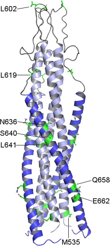
Three-dimensional representation of the trimeric gp41 protein ectodomain. Ribbon representation of the protein with the HR1 domain (positions 531–591) in light blue and the HR2 (positions 624–681) domain in blue. Positions relevant for the R5 or R5X4 tropism (see Fig. 2) are shown in green. The image was obtained from a consensus homology model generated with Prime software [49] from gp160 (Uniprot: Q70626, positions: 531–681) of HIV-1 group M subtype B (isolate LW123), and using two templates (PDB ID's: 2X7R and 1IF3) [47, 48]. The coordinates of this structure are available in the Additional file 4: Figure S1 (Gp41 coordinates - Homology model)