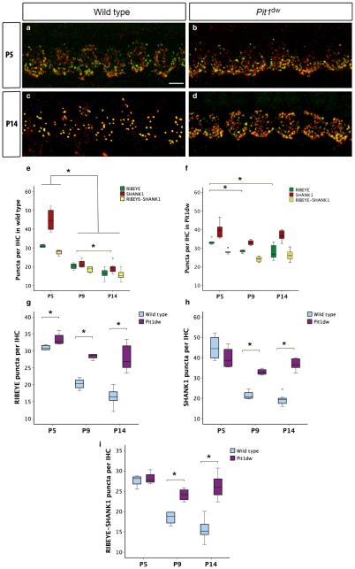
Fig. 1.
Synaptic pruning is disrupted in Pit1dw IHCs. (a and b) Projection of confocal sections obtained from the mid-turn of cochlear whole mounts stained with afferent presynaptic (RIBEYE; green) and postsynaptic (SHANK1; red) markers at P5 in WT mice (a) and Pit1dw mice (b). (c and d) The same markers at P14 in WT mice (c) and Pit1dw mice (d). (e–i) Box plots of the quantification of RIBEYE, SHANK1 and RIBEYE–SHANK1 puncta from the mid-turn of the cochlea in WT and Pit1dw mice. (e and f) RIBEYE, SHANK1 and RIBEYE–SHANK1 counts at P5, P9 and P14 in WT mice (e) and Pit1dw mice (f). (g–i) Comparison of RIBEYE (g), SHANK1 (h) and RIBEYE–SHANK1 (i) counts between WT and Pit1dw mice. significant comparisons (P < 0.05) are indicated with asterisks. The dots indicate outliers in the data.

