Fig. 6.
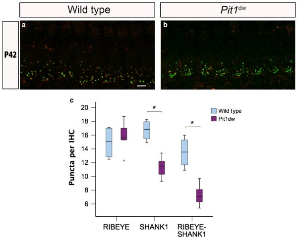
Reduced number of afferent postsynaptic counts in young adult Pit1dw IHCs. (a and b) Projection of confocal sections obtained from the mid-turn of cochlear whole mounts stained with RIBEYE (green) and SHANK1 (red) markers in WT mice (a) and Pit1dw mice (b) at P42. Scale bar: 10 lm. (c) Box plot of the quantification of RIBEYE, SHANK1 and RIBEYE–SHANK1 puncta from the mid-turn of WT and Pit1dw cochlea at P42. significant comparisons (P < 0.05) are indicated with asterisks. The dots indicate outliers in the data.

