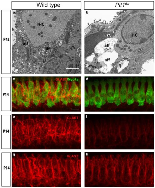
Fig. 7.
Swollen afferent type I terminals and reduced GLAST expression in young adult Pit1dw mice. (a and b) Representative TEM images from the mid-turn of Pit1dw and WT cochleas at P42. IHC and afferent boutons (aff) are indicated. Scale bar: 2 lm. (c and d) Projections of confocal sections obtained from the mid-turn of cochlear whole mounts co-stained with anti-GLAST (red) and anti-myosin VIIa (green) antibodies in WT and Pit1dw mice at P14. (e and f) GLAST expression (red) alone in WT and Pit1dw mice at P14 before TH treatment. (g and h) GLAST expression (red) at P14 in saline-treated WT mice and in TH-treated Pit1dw mice from P3 to P8. Scale bar: 10 lm.

