. 2015 Sep 28;7(1):234–239. doi: 10.1039/c5sc02857d

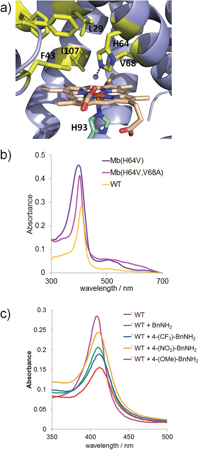
Fig. 1. (a) Distal pocket of sperm whale myoglobin (pdb 1A6K 12). Active site residues (yellow), heme (wheat), ‘proximal His’ (green), and heme-bound water molecule (grey sphere) are highlighted. (b) Electronic absorption spectra for wild-type Mb and Mb variants Mb(H64V) and Mb(H64V,V68A) in phosphate buffer at pH 7.0. The Soret bands have maxima at 408, 398, and 403 nm, respectively. (c) Ligand-induced decrease of Soret band intensity upon incubation of Mb in phosphate buffer at pH 7 with 20 mM benzyl amine (BnNH2) and para-substituted BnNH2 derivatives (λmax = 408 nm (WT); 409 nm (WT + BnNH2); 410 nm (WT + 4-(CF3)–BnNH2); 409 nm (WT + 4-(NO2)–BnNH2); 412 nm (WT + 4-(OMe)–BnNH2).

Fig. 1