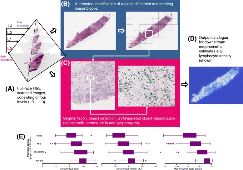
Fig. 1.

Overview of the image processing method. a Full-face H&E scanned images consist of four levels (L0–L3) across a gradation of resolutions. The levels L3 (lowest resolution) and L0 (highest resolution) are used to process each image. b Automated identification of regions of interest was performed by dividing image layer L3 into several small blocks (grid) and by analysing the pixel intensity distribution of each block. c Each image block found to contain tissue was mapped onto layer L0 and image segmentation and object detection (green ellipses) was conducted to construct an object catalogue. d Illustrative representation as a contour map of lymphocyte density derived using a k-nearest neighbour algorithm of the 50 nearest like-class neighbours. e Distribution of lymphocyte metrics by categories of lymphocytic infiltration based on central pathology review. SVM support vector machine
