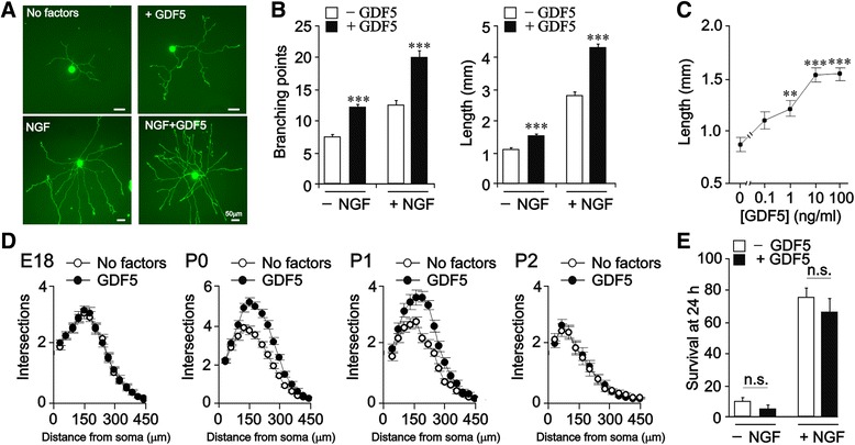
Fig. 1.

GDF5 promotes neurite growth from cultured neonatal SCG neurons. a Photomicrographs of representative P0 SCG neurons cultured for 24 h with or without 10 ng/ml GDF5 and/or 10 ng/ml NGF. b Branch point number and total length of neurite arbors of P0 SCG neurons cultured for 24 h with or without 10 ng/ml GDF5 and/or 10 ng/ml NGF. c Neurite arbor lengths of P0 SCG neurons cultured for 24 h with different concentrations of GDF5. d Sholl analysis of the arbors of E18, P0, P1 and P2 SCG neurons cultured for 24 h with and without 10 ng/ml GDF5. Cultures without NGF received 50 μM Boc_D_FMK to prevent apoptosis. Mean ± SEM of data from at least 150 neurons in each condition from 3 independent experiments are shown (** P < 0.01 and *** P < 0.001, statistical comparison with control, one-way ANOVA with Fisher’s post hoc). e Percentage survival of P0 SCG neurons cultured for 24 h in the absence of Boc_D_FMK with or without 10 ng/ml GDF5 in the absence or presence of 10 ng/ml NGF (mean ± SEM of the results of 3 independent experiments; n.s. = not significant)
