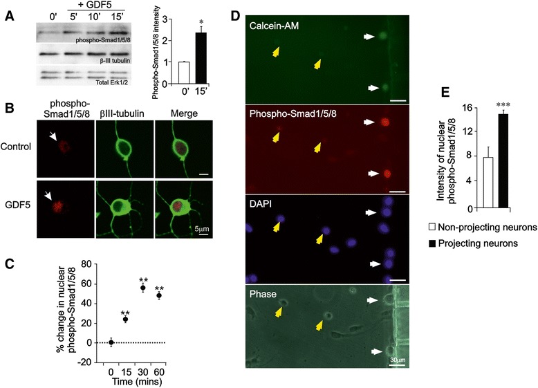
Fig. 4.

GDF5 promotes retrograde canonical Smad signalling along SCG axons. a Representative Western blot for phospho-Smad1/5/8 in P0 SCG neurons treated with GDF5 for 5, 10, and 15 min or untreated (0′) 12 h after plating using βIII tubulin and total ERK1/2 as loading standards. The bar chart plots the relative levels of phospho-Smad1/5/8 from densitometry of multiple blots at the 0 and 15 min time points (mean ± SEM). b Representative P0 SCG neurons immunolabeled for phospho-Smad-1/5/8 and β-III tubulin after either 30 min treatment with GDF5 or untreated (Control) 12 h after plating. c Percentage increase in nuclear phospho-Smad-1/5/8 immunolabeling in P0 SCG neurons treated with GDF5 for the indicated times relative to untreated control levels. d Representative P0 SCG neurons immunolabeled for phospho-Smad-1/5/8 after 60 min treatment of their axons with 10 ng/ml GDF5 in compartment cultures 12 h after plating. Addition of calcein-AM to the axon compartment was used to identify neurons that had projected axons into the axon compartment (white arrows), whereas unlabelled cells (yellow arrows) had not projected axons into the axon compartment. DAPI labelling indicates all cell nuclei in the field and the phase contrast image shows the location of the compartment barrier with two of its microchannels. Neurons whose axons had been exposed to GDF5 had a clear increase in nuclear accumulation of phospho-Smad proteins. Scale bar = 30 μm. e Relative intensity of nuclear phospho-Smad immunofluorescence in neurons with axons that had or had not projected into the axon compartment (mean ± SEM, *P < 0.05, ** P < 0.01, *** P < 0.001, statistical comparison with control, one-way ANOVA with Fisher’s post hoc)
