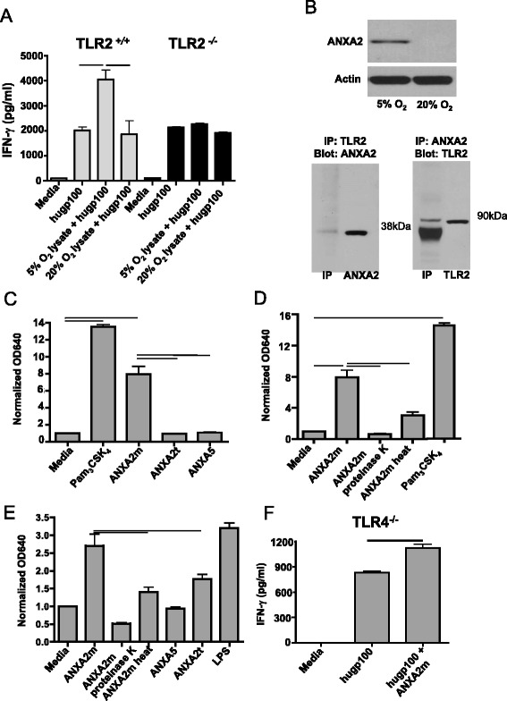
Fig. 1.

ANXA2 is enriched in physiologic oxygen, the monomeric form of which is a TLR2 agonist. a Splenocytes from B6 (TLR2 +/+) and TLR2 −/− mice were pulsed with the human gp10025–33 peptide, with or without lysates from GL261 cells grown under 5 % or 20 % O2. Addition of purified pmel-1 CD8 T cells resulted in INF-γ secretion, which was quantified after 48 h by flow cytometry. The experiment was repeated three times with similar results (the average INF-γ median fluorescence intensities [MFIs] for the three experiments were 1,868 and 4,057 for splenocytes stimulated with lysates from GL261 cells grown under 5 % or 20 % O2, respectively). Lines indicate p <0.05 by unpaired t-test. b (top) Protein in GL261 lysate was separated by SDS-PAGE and immunoblotted for murine ANXA2. (bottom) ANXA2 and TLR2 in GL261 lysates coimmunoprecipitate as shown by immunoblot. Recombinant murine ANXA2 (38 kDa) and TLR2 (90 kDa) were used as controls. c & d Compounds (6.9 μM) were pulsed onto human TLR2-expressing HEK-Blue cells, and secreted alkaline phosphatase was measured. “Proteinase K” and “heat” respectively indicate pre-treatment with the enzyme or incubation of the protein at 56 ° C for 30 min prior to addition to reporter cells. Y axes represent colorimetric quantification of alkaline phosphatase activity, measuring absorbance at 640 nm and normalized to media controls. The experiment was repeated three times with similar results. The mean absorbances at 640 nm for these three experiments were 7.2 and 1.4 for ANXA2m and ANXA2t, respectively. e Same as c and d only human TLR4-expressing HEK-Blue cells were used. The experiment was repeated three times with similar results. The mean absorbances at 640 nm for these three experiments were 2.7 and 1.7 for ANXA2m and ANXA2t, respectively. f Same as (a) except that splenocytes from TLR4 −/− mice were pulsed with the human gp10025–33 peptide with or without human ANXA2m. The experiment was repeated three times with similar results (the average INF-γ MFIs for the three experiments were 1,126 and 831 for peptide-pulsed splenocytes with or without human ANXA2m, respectively). Error bars are ± SEM, and lines show p <0.01 by unpaired t-test
