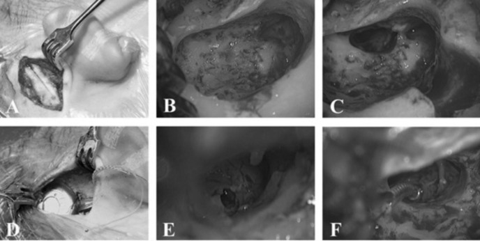
Fig. 1.
Intraoperative images of the surgical procedure. A: retroauricolar incision and subperiosteal dissection; B: “minimal“ mastoidectomy; C: posterior tympanotomy; D: insertion of receiver/stimulator in subperiosteal pocket after drilling of a well; E: iuxtafenestral cochleostomy; F: insertion of electrode array.

