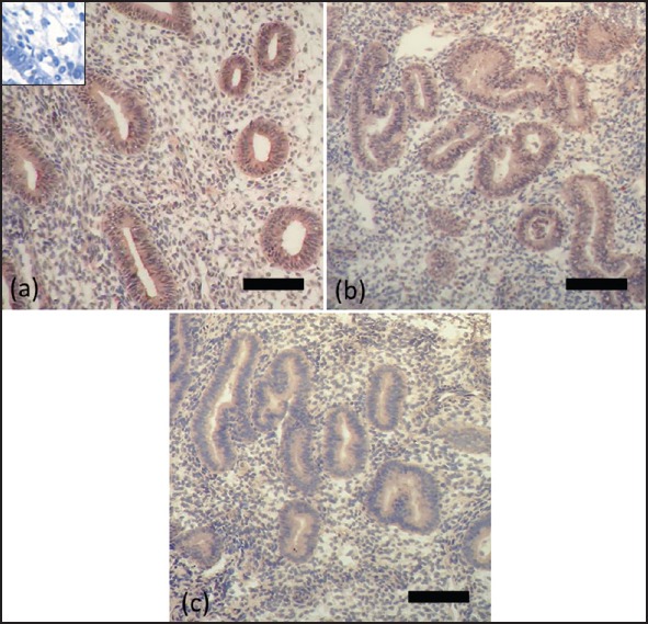
Figure 4.

Immunohistochemistry for apolipoprotein A1 using endometrial samples from PCOS patients (a) and normal women during proliferative phase (b) and secretory phase (c). Positive staining is brown and negative staining is blue. Insets show blocking of the antiapolipoprotein A1 antibody with its specific peptides. Bar = 50 μm
