-
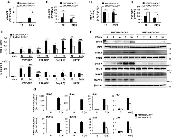
A, B
Virus replication at 12 and 24 hpi, in HDAC6
+/+ and HDAC6
−/− BMDMs in response to VSV‐GFP (MOI = 10) infection (A) and PR8‐GFP (MOI = 5) infection (B).
-
C
Virus replication in HDAC6
+/+ and HDAC6
−/− BMDMs in response to HSV‐GFP (MOI = 2) infection.
-
D
Virus replication in HDAC6
+/+ and HDAC6
−/− PBMCs in response to VSV‐GFP (MOI = 10) infection.
-
E
ELISA of IFN‐β (upper), IL‐6 (lower) levels in the supernatant of (A) and (B), and in HDAC6
+/+ and HDAC6
−/− BMDMs treated with poly(I:C) (20 μg/ml) or transfected with 5′ppp‐dsRNA (1 μg/ml).
-
F
Immunoblot analysis of the phosphorylated and inactive forms of IRF3, IKBα, TBK1, RIG‐I, MAVS, HDAC6, and β‐actin at the indicated times (0, 2, 4, 8, and 16 h) in HDAC6
+/+ and HDAC6
−/− BMDMs. BMDMs were stimulated with PR8‐GFP (MOI = 3).
-
G
Induction of mRNA for type I IFN, IL‐6, and other IFN‐related antiviral genes in HDAC6
+/+ and HDAC6
−/− BMDMs in response to a RIG‐I agonist stimulation at 6 h. HDAC6
+/+ and HDAC6
−/− BMDMs were stimulated with 5′ppp‐dsRNA (0.5 μg/ml) for 6 h.
Data information: Data are representative of at least two independent experiments. Error bars, mean ± SD. *
‐test).