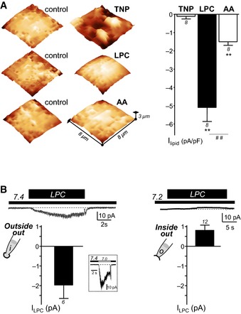
Figure 4. Effect of trinitrophenol (TNP), LPC, and AA on the cell membrane shape and ASIC3 channel activity at pH 7.4.

- Representative SICM experiments (left panel) performed on HEK293 cells showing the effects of the crenator trinitrophenol (TNP), of LPC (bovine brain extract) and of AA on the membrane shape (TNP at 5 mM, LPC at 30 μM and AA at 10 μM). SICM images are obtained before (control) and after extracellular application of TNP, LPC, or AA. Whole‐cell recording experiments (right panel), performed at −80 mV, showing the corresponding effect of TNP (5 mM, 30‐s applications), LPC (10 μM, 10‐s applications), and AA (10 μM, 30–s applications) on rat ASIC3 channels at resting pH 7.4 (**P < 0.01, significant current induced at pH 7.4, and ## P < 0.01, Wilcoxon tests).
- Effect of LPC applied either intracellularly or extracellularly in excised patch‐clamp experiments recorded from rat ASIC3‐transfected F‐11 cells. Outside‐out (left panel) and inside‐out (right panel) currents were recorded at −50 mV and +50 mV, respectively. Histograms show the mean amplitudes of the currents induced by LPC (10 μM) applied either at pH 7.4 (extracellular side, outside‐out) or at pH 7.2 (intracellular side, inside‐out). These two different pH values mimic the resting physiological pH of extracellular and intracellular media, respectively. For outside‐out experiments, LPC‐induced current amplitude is measured from patches that also displayed a typical ASIC3 inward current following extracellular acidification from pH 7.4 to pH 7.0 (inset). The dotted lines represent the basal current level.
