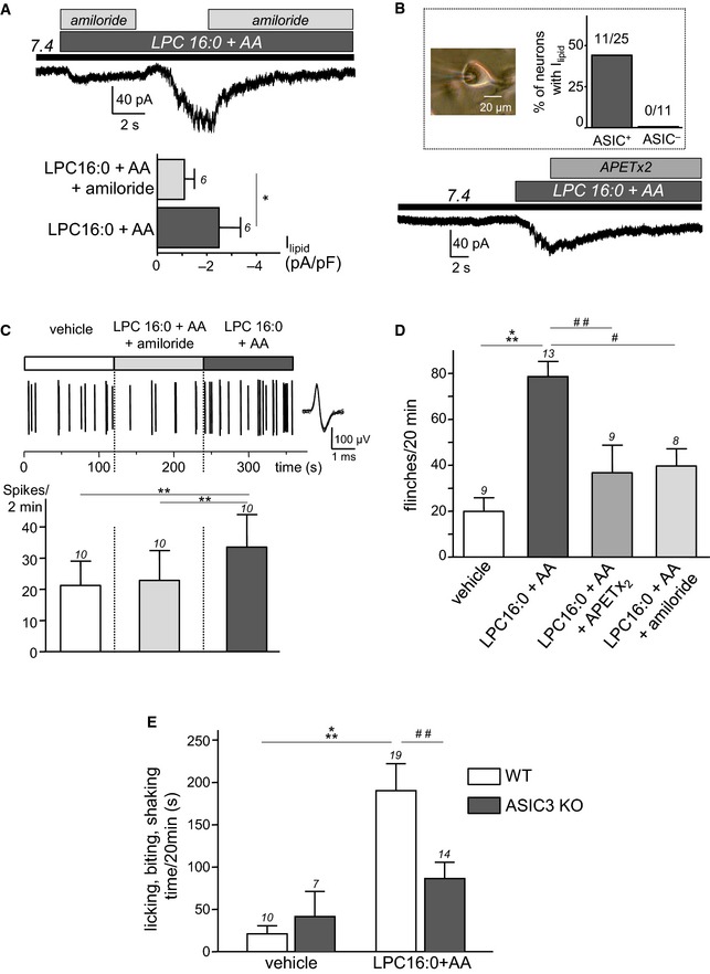
Effect of amiloride (1 mM) on native whole‐cell currents recorded at −80 mV and induced by the co‐application of LPC and AA (10 μM each) at pH 7.4 on rat DRG neurons (*P < 0.05, Wilcoxon test).
Typical inhibitory effect of APETx2 (1 μM) on the lipid‐induced (LPC16:0 + AA) current recorded at pH 7.4 from rat DRG neurons. Inset: percentage of DRG neurons (picture, scale bar = 20 μm) responding to lipids and that displayed (ASIC+) or do not displayed (ASIC−) a pH 6.6‐induced ASIC current.
Nerve‐skin experiments showing the effect of lipids (LPC16:0 + AA, 48 nmole each), applied for 2 min onto the receptive fields of C‐fibers in the skin, either with amiloride (1 mM) or alone. Top trace shows a typical recording of C‐fiber firing with an enlarged action potential (AP) on the right, while the average spike frequency is represented at the bottom. Lipids provoke an increase of AP firing, which is blocked when co‐applied with amiloride (**P < 0.01 for LPC16:0 + AA vs. vehicle and LPC16:0 + AA vs. LPC16:0 + AA + amiloride, Friedman test followed by a Dunn's post hoc test).
Pain behavioral experiments showing the number of rat flinches induced by a hindpaw injection of LPC16:0 + AA (2.4 nmole each) or vehicle (0.24% ethanol). Lipids are injected either alone or in the presence of APETx2 (0.2 nmole) or amiloride (20 nmole) (#
P < 0.05, ##
P < 0.01 and ***P < 0.001, Kruskal–Wallis test followed by Dunn's post hoc tests).
Lipid‐induced pain in wild‐type (WT) and ASIC3 knockout mice (ASIC3 KO). Lipids (LPC16:0 + AA, 4.8 nmole each) or vehicles (0.96% ethanol) are injected into a hindpaw, and the pain behavior is represented as the time spent by mice licking, biting, and shaking their injected paw for 20 min (##
P < 0.01 and ***P < 0.001, two‐way ANOVA tests followed by Bonferroni's post hoc tests).
) is indicated right of or above each bar graph, and error bars represent ± SEM.

