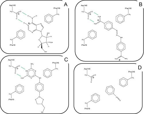
Figure 7.

Schematic model for the interactions between TbAT1 ligands adenosine (A), diminazene (B) and melarsoprol (C) with the critical amino acid residues in the TbAT1 binding pocket; Frame (D) shows the putative interaction of phenylarsine oxide with the binding pocket. Hydrogen bonds are depicted as dotted lines. The image was created using ChemDraw Pro 10.0, CambridgeSoft.
