
Figure 2.
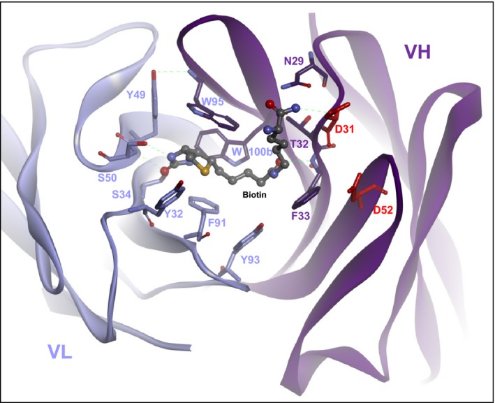
Structure of an antibody that specifically binds conjugated biotin (biocytinamide in this figure) but not free biotin ( PDB 4S1D). Note the negatively charged aspartate 31 and aspartate 52 of the VH domain lining the sides of the binding pocket. The negative charges of their side chains generate a charge repulsion which prevents entry of (COOH‐containing) free biotin. In contrast, biotin that is coupled to a payload and thereby is uncharged (in the same manner as biotcytinamide that was used to solve the structure) can enter and stay in the binding pocket without charge repulsion.