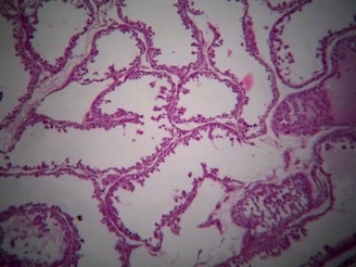
Figure 3.

General testicular degeneration with a widespread tubular atrophy and a significant decrease of the testicular parenchyma. Seminiferous tubules were covered by one or two rows of cells with epithelial vacuolization. The basal membrane appeared with slightly reduced diameter and irregular profile. Sertoli cells appeared normal, while there was a complete lack of spermatogonia
