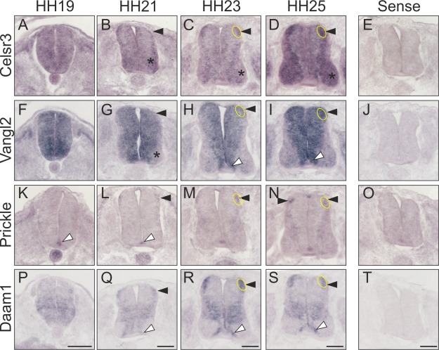
Figure 1.

PCP pathway components are expressed in chicken dI1 commissural neurons. Transverse sections of spinal cords taken from chicken embryos at the indicated developmental stages were subjected to in situ hybridization. (A–D) Celsr3 was widely expressed in the neural tube at HH19 (A). By HH21, Celsr3 expression was mainly found in dI1 neurons (arrowhead), as well as in more ventral populations of interneurons and motoneurons (asterisk). The expression pattern was largely maintained at HH23 (C) and HH25 (D). (E) No staining was seen after hybridization with the sense probe derived from Celsr3. (F–I) Vangl2 mRNA was found throughout the neural tube at HH19 (F). A decrease of Vangl2 expression was found in motoneurons (asterisk) and mature interneurons, except dI1 neurons (arrowhead), at HH21 (G). Vangl2 expression was maintained in dI1 neurons (arrowhead) at HH23 (H) and HH25 (I). Expression of Vangl2 was also seen in the floor plate and in cells adjacent to the floor plate (white arrowhead). (J) No signal was seen with the Vangl2 sense probe. (K–N) In contrast to the cell surface molecules, the distribution of the intracellular components of the PCP pathway was much more restricted during the time window of commissural axon guidance. Prickle was found in the floor plate already at HH19 (white arrowhead, K). By HH21, Prickle expression was detectable also in dI1 neurons (arrowhead, L). Expression in dI1 neurons persisted at HH23 (M) and HH25 (N). (O) No staining was seen with a Prickle sense probe. (P–S) Daam1 was found in dI1 neurons at all stages (arrowhead). In addition, expression of Daam1 was found in cells adjacent to the floor plate (white arrowhead). (T) No staining was seen with a Daam1 sense probe. The location of dI1 commissural neurons is indicated. Scale bars: 100 µm. [Color figure can be viewed in the online issue, which is available at wileyonlinelibrary.com.]
