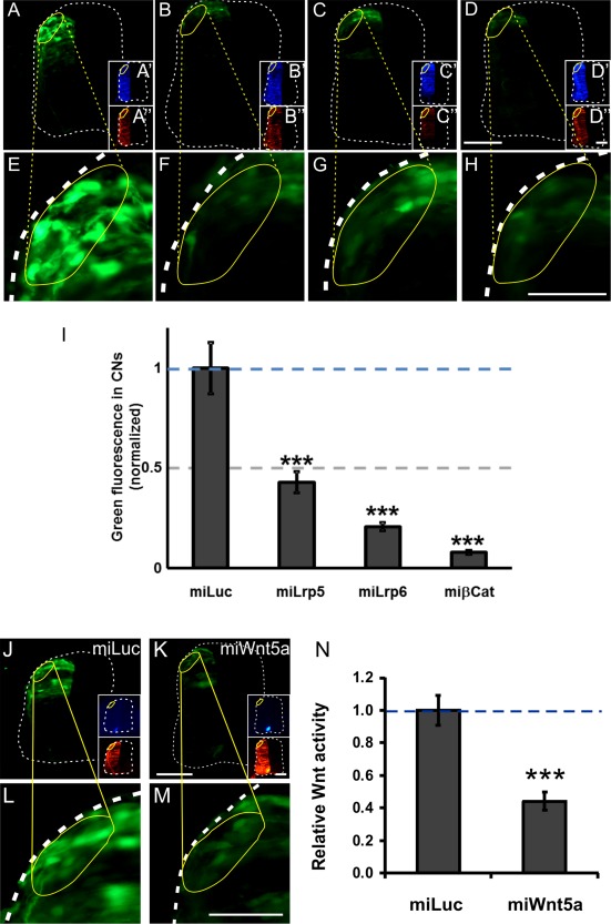
Figure 7.

Canonical Wnt signaling persists in mature dI1 commissural neurons. Canonical Wnt signaling in mature dI1 neurons at the time when their axons crossed the floor plate and turned into the longitudinal axis was demonstrated by expression of GFP under the control of a TEF/Lef‐responsive promoter (A,E,I). Signaling was efficiently blocked after silencing Lrp5 (B,F,I), Lrp6 (C,G,I), or β‐Catenin (D,H,I) with miRNAs. Inserts (A'–D') show EBFP2 (enhanced blue fluorescent protein) expression indicating the successful electroporation of the miRNA constructs. Tomato fluorescence was used to normalize transfection efficiency (A“–D”). The relative GFP expression (ratio GFP/Tomato) seen in miLuc‐transfected embryos was set to 1.0 (I; 1.0 ± 0.13; 4 embryos, 27 sections). Injection and electroporation of miLrp5 reduced relative GFP expression to 0.43 ± 0.05 (4 embryos, 29 sections). Electroporation of miLrp6 resulted in GFP values of 0.21 ± 0.02 (4 embryos, 31 sections), and targeting β‐Catenin reduced GFP to 0.08 ± 0.01 (4 embryos, 31 sections). One‐way ANOVA was used for statistical analysis *** p < 0.001. Similarly, silencing Wnt5a in the floor plate resulted in a reduction of canonical Wnt signaling in dI1 neurons in vivo (J–N). Baseline canonical Wnt activity was calculated from the ratio between GFP and Tomato fluorescence in miLuc‐treated embryos (J,L; set to 1.0). After silencing Wnt5a in the floor plate (see EBFP2 expression to visualize effective targeting of the floor plate) canonical Wnt signaling was efficiently reduced (K,M,N). (N) Quantification of green fluorescence normalized by red fluorescence. t‐test, *** p < 0.001. Scale bars: 50 µm. [Color figure can be viewed in the online issue, which is available at wileyonlinelibrary.com.]
