Figure 3.

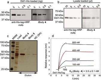
Application of iBody 4, which targets His‐tagged proteins. a) Comparison of iBody 4 and anti‐polyhistidine antibody sensitivity for the visualization of purified Ddi1‐His by western blot. b) Comparison of iBody 4 and the anti‐polyhistidine antibody for the visualization of Ddi1‐His in a cell lysate by western blot. c) Affinity isolation of Ddi1‐His by using iBody 4. iBody 5 (which does not possess the tris‐NTA ligand), and blank resin (NC) were used as negative controls. MWM=molecular‐weight marker. d) Binding of Ddi1‐His to immobilized iBody 4 as analyzed by SPR.
