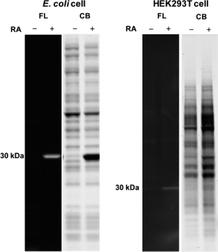
Figure 6.
Selectivity of P1 in E. coli or HEK293T cell lysate lacking or overexpressing RA. Lysates (3 mg/mL) obtained by sonication were incubated with P1 (10 μM) for 10 min at 25 °C. The samples were fractionated on an SDS-PAGE gel and visualized by either a Bio-Rad Gel Doc Imager employing UV illumination to see the conjugate fluorescence signal or bright field light to observe the Coomassie staining. No significant off-target bands were observed in lysates of cells lacking or expressing RA. FL = RA-P1 conjugate-associated fluorescence, CB = Coomassie blue.

