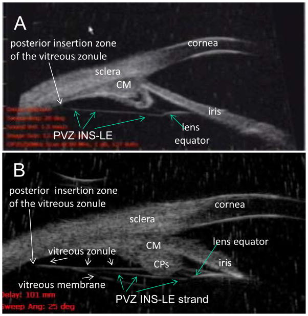
Figure 1.
(Panel A) Ultrasound biomicroscopy (UBM) images (50 mHz) in the nasal quadrant of a 19-year-old male human. There was always a prominent continuous strand that extended from the posterior insertion zone of the vitreous zonule directly toward the posterior aspect of the lens equator and attached to it, seen in all 12 human subjects and termed PVZ INS-LE strand (which stands for posterior vitreous zonule insertion to lens equator strand). (Panel B) UBM image in a 65-year-old human subject. Note that a portion of the PVZ INS-LE strand is visible that extends between the posterior insertion zone of the vitreous zonule and the posterior lens equator, as in (A) with the strand passing immediately internal to the CPs. The PVZ INS-LE strand lies between the vitreous zonule and vitreous membrane. Note: Panel B shows both the vitreous zonule and the PVZ INS-LE strand. During accommodation the muscle pulls forward the vitreous zonule and the PVZ INS-LE strand. The PVZ INS-LE strand does not relax during accommodation. [Croft, Kaufman and Lütjen-Drecoll: ARVO 2010 IOVS 2013] CM = ciliary muscle. Reprinted with permission from: Croft et al. Accommodative Movements of the Vitreous Membrane, Choroid, and Sclera in Young and Presbyopic Human and Nonhuman Primate Eyes. Invest Ophthalmol Vis Sci 2013;54:5049–5058; copyright Association for Research in Vision and Ophthalmology (ARVO).

