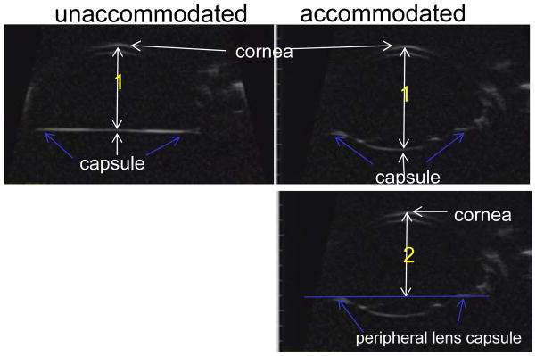
Figure 3.
Ultrasound biomicroscopy (UBM) images taken in a monkey eye following extracapsular lens extraction (ECLE). UBM allows visualization of the anterior chamber and capsule following ECLE. The following distances were measured: Distance 1: The axial distance from the posterior central capsule to the corneal epithelium was measured in the resting and accommodated states. Distance 2: The axial distance from the peripheral lens capsule (blue arrows) to the corneal epithelium was measured in the resting and accommodated states.

