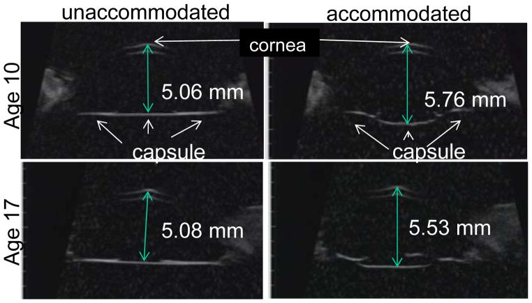
Figure 8.
Ultrasound biomicroscopy (UBM) images obtained in rhesus monkey eyes two weeks after ECLE in the resting and maximally accommodated states. Numbers represent the axial distance of the central capsule from the cornea. During accommodation the central capsule bows backward, but not the peripheral capsule. The accommodative backward bowing is diminished with age.

