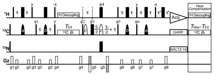
Figure 1.
2D 13C CEST pulse sequence for characterizing slow chemical exchange in nucleic acids. Narrow (wide) rectangles are 90° (180°) pulses and closed (open) shapes are selective on (off) resonance 180° pulses. Delays are τ = 1/4JCH and τ′ = g8. Phase cycle is ϕ1 = {x,−x}, ϕ2 = {y}, ϕ3 = {2x,2y,2(−x),2(−y)}, ϕ4 = {4x,4(−x)}, ϕ5 = {4x,4(−x)}, ϕ6 = {4y,4(−y)}, receiver = {x,2(−x),x,−x,2(x),−x}. Briefly, 1H magnetization is transferred to 13C longitudinal magnetization, which relaxes under a weak 13C B1 field during TEX. 13C transverse magnetization then evolves during t1 and is returned to 1H for detection. Peak intensities are monitored as a function of B1 offset and power. See details in SI.

