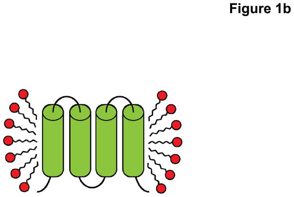
Fig. 1.
Cartoon representation cross-section of a membrane protein solubilized in (a) lipid bilayer of the host cell and (b) detergent introduced during the purification process. Upon removal of the protein from the native lipid (blue) environment, the protein-detergent complex consists of a uniform disordered ring of detergent monomers (red) making hydrophobic contacts with the surface of the protein (green).


