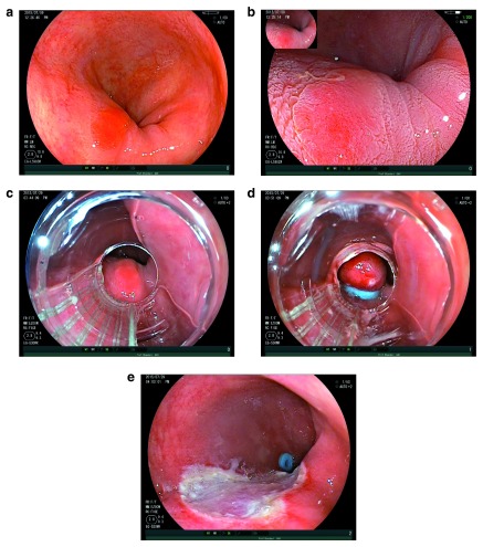
Figure 2. EMR of Barrett’s HGD.
( a) An area of Barrett’s high-grade dysplasia. ( b) The same area demonstrating acetowhitening effect. ( c) The same lesion as viewed down a multi-band ligator. ( d) Pseudopolyp created by the band ligator. ( e) Resection defect following endoscopic mucosal resection.

