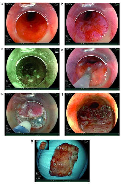
Figure 3. ESD of Barrett’s IMC.
( a) pT1a/M3 intramucosal cancer arising in Barrett’s oesophagus. ( b) The same lesion following acetic acid. Note the differential early loss of acetowhitening. ( c) Edges of the lesion marked with endoscopic submucosal dissection (ESD) knife under virtual chromoendoscopy. ( d) Submucosal injection. ( e) Mucosal incision with ESD knife. ( f) Resection base following ESD. ( g) Resection specimen.

