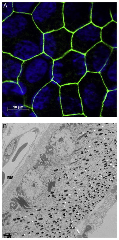
Figure 4.

(A) Cultured human fetal RPE cells. Tight junctions, shown stained for ZO-1 (zonula occludens-1, in green), ensure that the RPE functions as an effective barrier between the retina and the choroid, with exchange tightly regulated through ion channels and transporters. (B) Transmission electron micrographs of chick RPE; cells show a distinct asymmetry, with nuclei located in the basal region adjacent Bruch’s membrane (BM) and choriocapillaris (CC), and melanin granules more concentrated in the apical (retinal) region.
