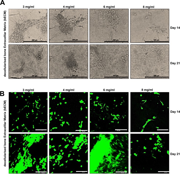
Fig 2. Cell morphology and viability of DPSCs in bECM scaffolds in vitro.
(A) Representative images of DPSCs seeded on bECM scaffolds (3, 4, 6, 8 mg/ml) at day 14 and 21. DPSCs proliferate, elongate and form clusters, scale bar: 100 μm. (B) DPSCs remained viable after 14 and 21 days of culture on 3, 4, 6, 8 mg/ml bECM hydrogel scaffolds. Viable cells stained green and dead cells stained red after CMFDA/PI staining. Cells were observed under fluorescence confocal microscope, scale bar: 100 μm.

