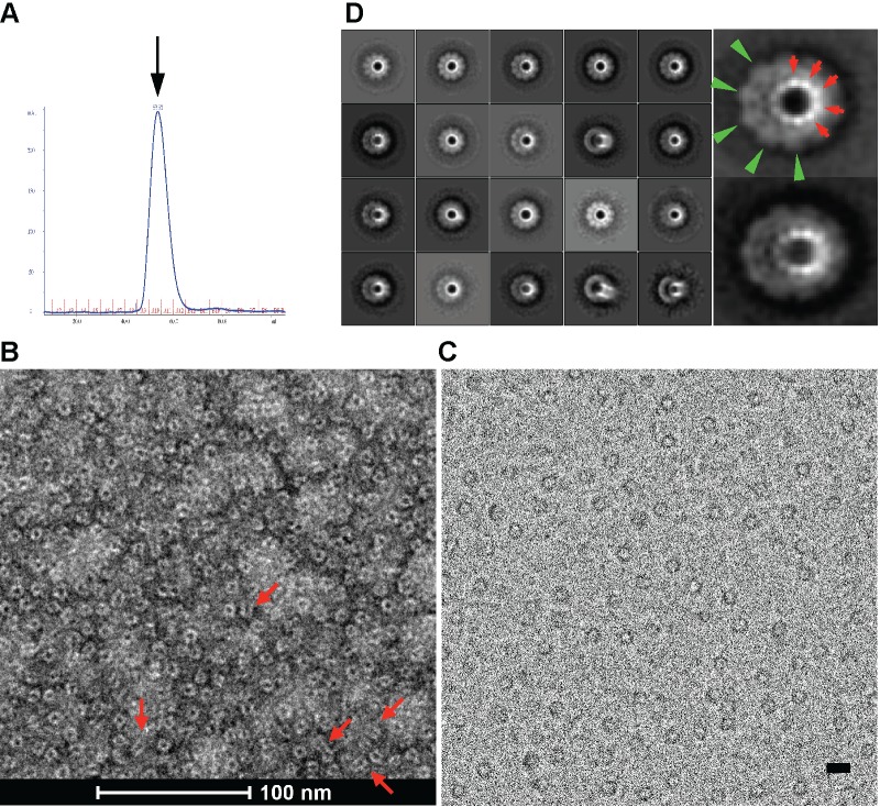
Fig 1. The gp12 assembles into a ring-like decamer.
(A) Size exclusion chromatogram of the purified gp12 decamer. The peak corresponds to an estimated molecular weight of 471kDa, indicating a decamer (arrow). (B) Electron micrograph of the purified gp12 decamer negatively stained with uranyl acetate. Notice the dominant views are down the longitudinal axis of the gp12 decamer. Some side or tilted views are indicated with red arrows. (C) Electron micrograph of the frozen-hydrated gp12 decamer. Bar, 100 Å. (D) Class averages of the frozen-hydrated gp12 decamer. Enlarged views of two class averages are shown on the right. The 10-fold symmetry is clearly evident for both the crown (green arrowheads) and the stem domains (red arrows). The box size for class averages in the left panel is 309.8 Å.

