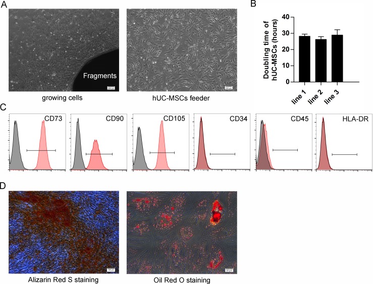
Fig 1. Characterization of hUC-MSCs.
(A) Growing cells from fragments (left) and MMC-treated hUC-MSCs feeder (right) show property of fibroblast-like cells with a spindle-shaped morphology. Scale bar: 200 μm. (B) The doubling time of different hUC-MSC lines. Each column represented the mean ±SD. (C) Flow cytometry results of rapidly dividing hUC-MSCs show they are positive for MSCs-specific markers (CD73, CD90 and CD105) but negative for CD34, CD45 and HLA-DR. (D) Alizarin Red S staining of osteogenic cells differentiated from hUC-MSCs (left). Scale bar: 100 μm. Oil Red O staining of adipogenic cells differentiated from hUC-MSCs (right). Scale bar: 20 μm.

