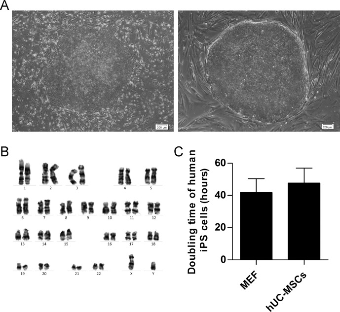
Fig 3. Morphology, Karyotype analysis and doubling time of human iPS cells.
(A) hiPSC colonies grown on MEF (left) and hUC-MSC (right) feeders at passage X+31 revealed undifferentiated hESC morphology with high nucleus/cytoplasm ratio. Scale bar: 200 μm. (B) G-band staining showed that hiPSC cells on hUC-MSCs feeder layers at passage X+31 maintained a normal karyotype. (C) The doubling time of hiPSCs cocultured on different feeders at passage X+31. Each column represented the mean ±SD.

