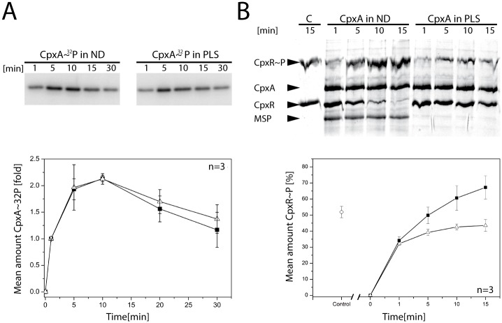
Fig 3. CpxA is functional in nanodiscs.
(A) Phosphorylation activities of the sensor histidine kinase CpxA in ND and PLS. Autophosphorylation was initiated by addition of [γ32-P]ATP. Samples were withdrawn at the indicated time points. The plot (lower panel) shows the results of densitometric analysis (of triplicate gels) of the autoradiograms in (A) The amount of CpxA~32P seen after 1 min was set to 1 and used for normalization of the other values. (B) Phosphotransfer from CpxA to CpxR. CpxR was phosphorylated using CpxA in ND (■) and in PLS (ρ), respectively, and a representative gel is shown (the experiment was performed in triplicate). A control experiment (lane marked with C; ◇ in the plot) was carried out by phosphorylating CpxR with acetyl-phosphate for 15 min. Phosphotransfer was initiated by concomitant addition of CpxR and ATP. Samples were withdrawn at the indicated time points and subjected to a Zn2+-Phos-tag™ PAGE. Densitometric analysis of the CpxR~P bands was performed and mean values were plotted (lower panel). Error bars indicate the standard deviation.

