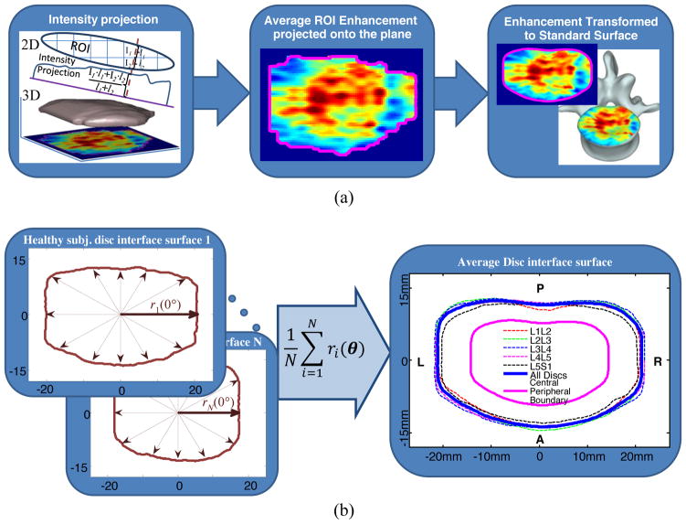
Fig. 4.
(a) Projection of voxel enhancement values onto a caudal plane and registration onto the template surface and mapping onto a representative vertebral body. (b) Schematic illustration of template surface generation using individual ROIs from healthy discs. The central region is shown with the magenta line and the peripheral region is the area between the magenta and the blue lines.

