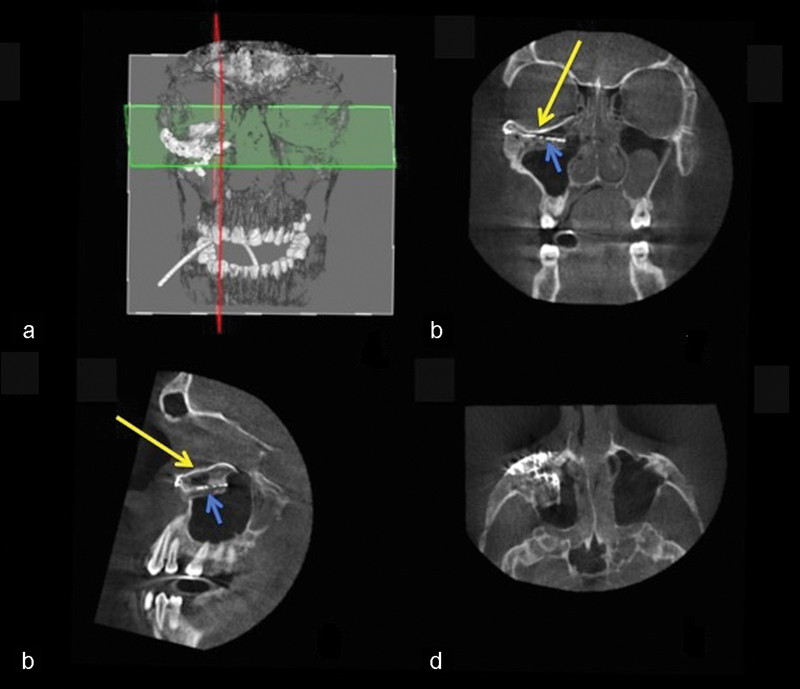
Fig. 12.

Case 3: Intraoperative 3D imaging using the Pulsera fluoroscopy system. (a) 3D reconstruction and section plane indication. (b) Good fit of the two puzzle pieces (long yellow arrow). Malpositioned mesh (short blue arrow). (c) Good fit of the medial puzzle-piece (long yellow arrow). The titanium mesh was not prebent and was 7 mm too low at the posterior sigmoid bulge (short blue arrow). (d) Axial view of the titanium mesh. The two-piece puzzle implants are visible only where they overlap the infraorbital rim.
